Mallory Trichrome kit
Product Description
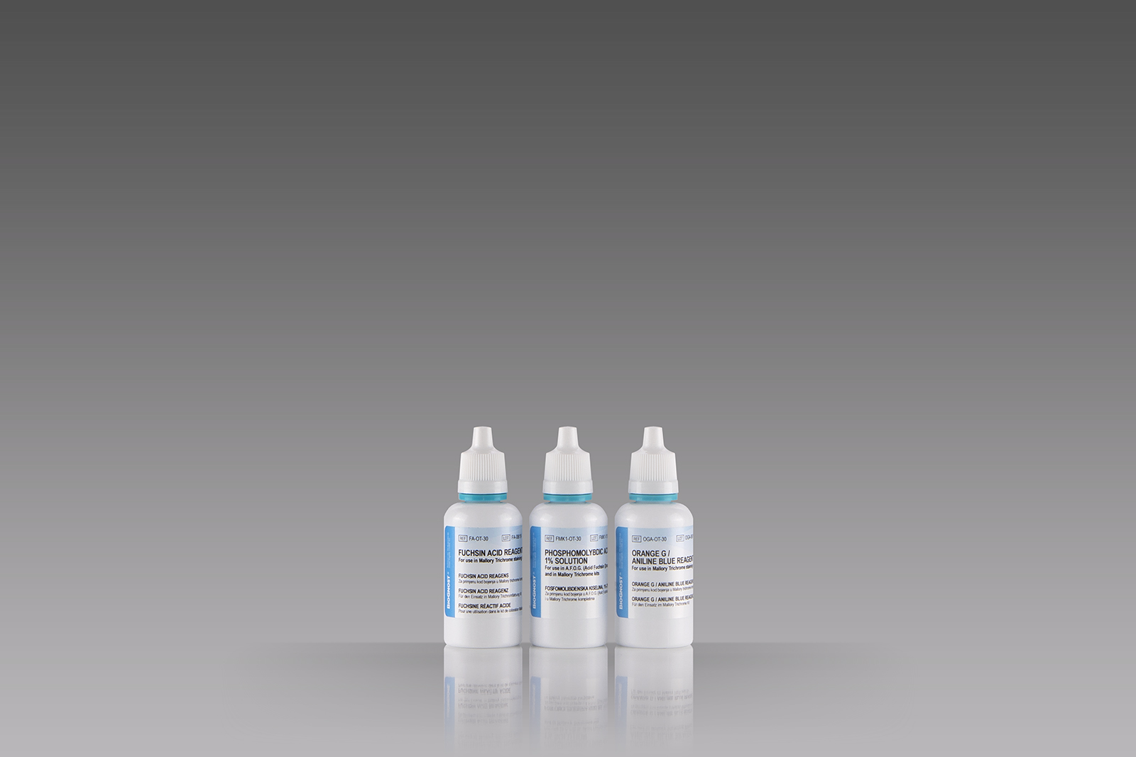
Three-reagent staining kit for connective tissue visualization and detection of collagen, cartilage, muscle, elastic fibers, mucous, pituarity cells, reticulum, bones, amyloid and erythrocytes.
Available Options:
Introduction
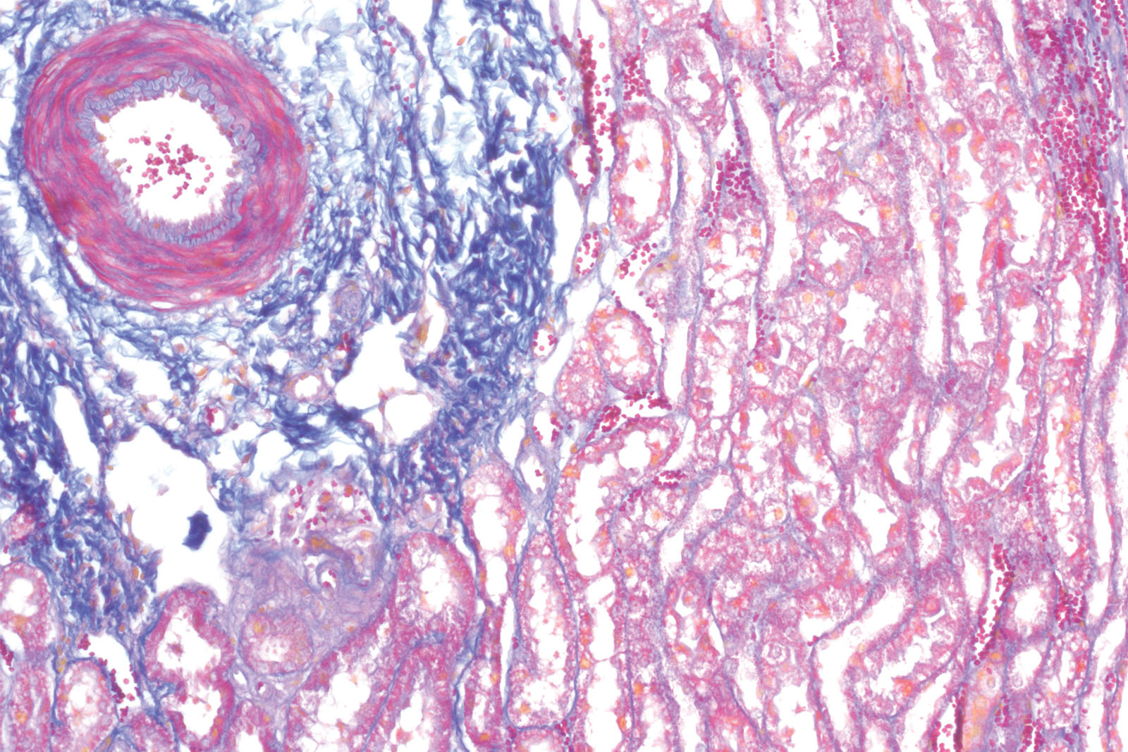
Mallory trichrome staining kit is used for treating the tested microscopic sample using three different stainings with differential counterstaining of two basic parts of the tissue (muscle and collagen fibers) in focus. By staining the sample with Fuchsin Acid acidic dye nuclei and muscles are stained red to pink. The phosphomolybdic acid molecule then pushes out Fuchsin Acid dye molecules from collagen and thus enables Aniline Blue to bind, resulting in collagen being stained contrast blue instead of red. Orange G (molecule of the lowest molar mass) stains erythrocytes.
Technical Data
Trade name: MALLORY TRICHROME KIT
Chemical name:
Catalogue number: MT-X**
Available volumes:
for 100 tests
3x100 mL
Storing, stability and expiry date:
Transport information:
Transporting/shipment by road (ADR) - Not classified
Transporting/shipment by sea (IMDG) - Not classified
Transporting/shipment by air (ICAO-TI/IATA-DGR) - Not classified


