Reticulin Contrast kit
Product Description
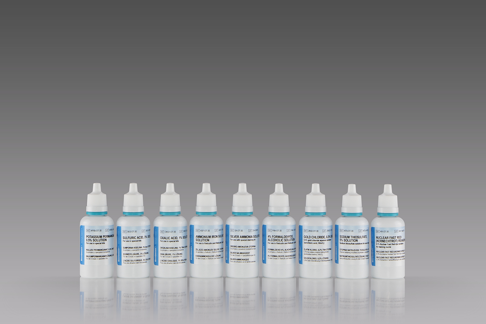
Nine-reagent kit for detecting argyrophilic reticulin fibers according to Gordon and Sweets. The kit contains gold chloride solution that enhances visualization of reticulin fibers and it also contains Nuclear Fast Red (Kernechtrot) reagent that enables fine contrasting background.
Available Options:
Introduction
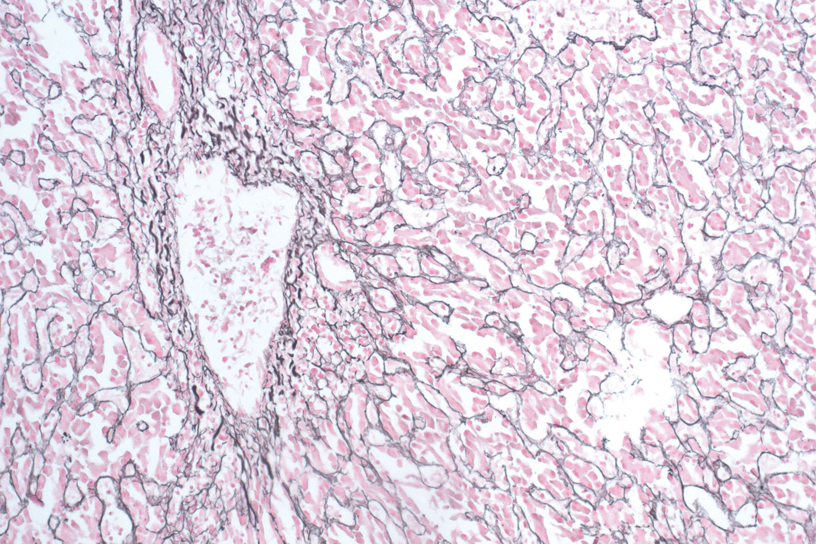
Reticulin Contrast kit is used for identification and easier visualization of argentaffin reticular fibers in connective tissue. Reticulin provides structural support. It is found in the liver, spleen and kidneys. Reticulin fibers are clearly defined in the healthy liver; necrotic and cirrhotic liver has discontinuous fibers. The visualization is based on silver depositions on reticulin fibers. The tissue sample must be oxidized with potassium permanganate. Silver is formed from ammonia solution containing silver nitrate and is deposited in the form of brown sediment on reticulin fibers. Formalin acts as a reducing agent and accelerates the procedure. Unbound silver is washed away using sodium thiosulfate. Reticulin Contrast kit also contains a gold chloride solution that stabilizes and tones the section’s image. The kit contains Nuclear Fast Red (Kernechtrot) counterstain.
Technical Data
Trade name: RETICULIN CONTRAST KIT
Chemical name:
Catalogue number: RET-X**
Available volumes:
for 100 tests
9x50 mL
9x100 mL
Storing, stability and expiry date:
Transport information:
Transporting/shipment by road (ADR) - Not classified
Transporting/shipment by sea (IMDG) - Not classified
Transporting/shipment by air (ICAO-TI/IATA-DGR) - Not classified


